
Advanced eye care, closer to you across Delhi NCR Appointments: +91 98991 18030
Diagnostics & Imaging
CONDITIONS & TREATMENTS

Eye Diagnostics & Imaging in Delhi NCR

Get a detailed picture of eye health with OCT, fundus imaging, corneal topography and visual field testing — supporting better decisions across every specialty.

OCTRetinal & anterior segment
FundusWidefield imaging
TopoCorneal mapping
HFAVisual field analysis
COMPARE YOUR OPTIONS

Diagnostics & Imaging — types & approaches

Each option has its own strengths. Your specialist will match the right pathway to your eyes and lifestyle.

01

OCT (Optical Coherence Tomography)

High-resolution cross-sections of the retina and cornea.

Essential in retina, glaucoma and refractive planning.
02

Fundus imaging

Documented photos of the back of the eye.

Supports diabetic and long-term retinal follow-up.
03

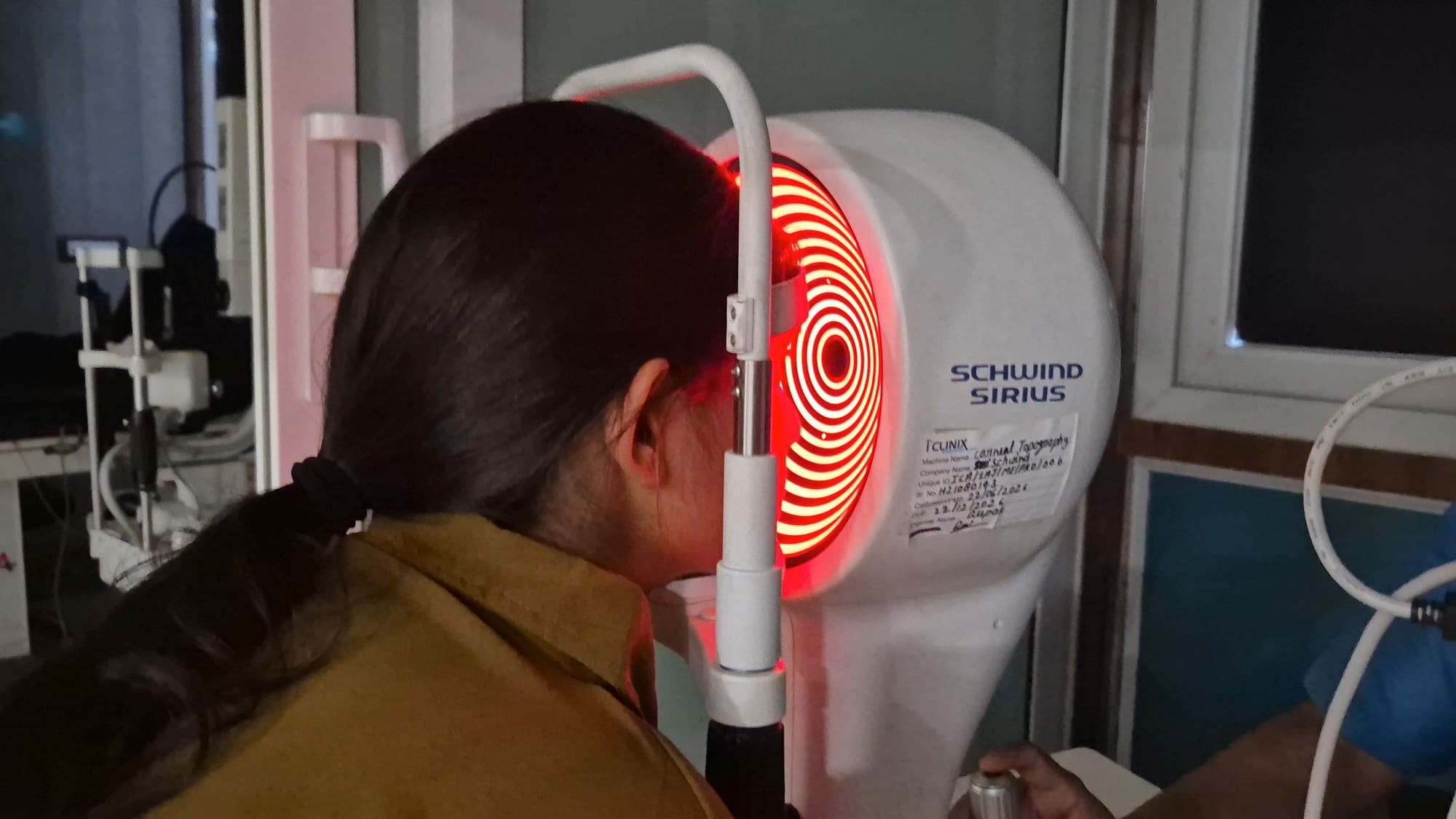
Corneal topography

Detailed corneal shape mapping.

Critical for refractive planning and keratoconus screening.
04

Visual field testing

Peripheral vision mapping.

Central to glaucoma diagnosis and follow-up.
COMPARE YOUR OPTIONS

Ocular angiography: FFA, ICG & OCTA

Angiography maps blood flow at the back of the eye — leakage, blockage and abnormal vessels a standard exam can miss.

01

FFA

Fluorescein dye, ~10–15 min.

Diabetic and vein disease, macular degeneration.
02

ICG

Indocyanine green dye, ~20–30 min.

Choroidal disease, central serous.
03

OCTA

No dye, seconds to minutes.

Fast, repeatable monitoring.
FFA/ICG: skin or urine may look yellow for up to a day
Mild nausea possible; allergic reactions are rare
OCTA covers a smaller area and can be affected by eye movement
Tell us about any kidney problems or dye allergies beforehand
ELIGIBILITY

Who is a good candidate?

Eligibility is confirmed through a detailed screening — the checklist below is a starting point.

Referred for a specialist opinion

Diabetic or hypertensive with no recent imaging

High myopia or family history of retinal disease

Pre-operative LASIK / cataract planning

Suspected glaucoma or optic nerve concerns

THE PROCEDURE

A clear, step-by-step pathway

01

Booking

Choose a convenient centre and time.

02

Imaging

Non-invasive imaging by trained optometrists.

03

Specialist review

Your treating doctor interprets results in context.

04

Plan

Understand what the findings mean for your care.

TECHNOLOGY

Modern platforms, careful decisions

Spectral-domain OCT
Widefield fundus camera
Corneal topography & tomography
Humphrey visual field analyser
YOUR SURGEONS

Specialists who treat diagnostics & imaging

COST & INSURANCE

Understanding cost, technology and recovery

Final cost depends on your eye, the recommended pathway, chosen technology and any lens or implant options. Our care team explains each factor before you decide, including cashless insurance and EMI where applicable.

  • Transparent estimates after screening
  • Cashless support across major TPAs
  • Structured recovery and follow-up plan
  • Second-opinion friendly consultations
Insurance & empanelments
AVAILABLE AT

Centres offering diagnostics & imaging

PATIENT QUESTIONS

Diagnostics & Imaging — patient questions

Is OCT safe?

OCT uses light — it is non-invasive and safe for repeat use, including in children.

Do I need imaging at every visit?

Only when clinically indicated — your specialist will explain.

Can I get imaging without a doctor referral?

Yes, but interpretation is best done together with a specialist consultation.

How long do the tests take?

Most scans take a few minutes each — the full workflow is typically under 30 minutes.

RELATED CARE

You may also want to read

YOUR NEXT STEP

Good eye care starts
with a conversation.

Call WhatsApp Book